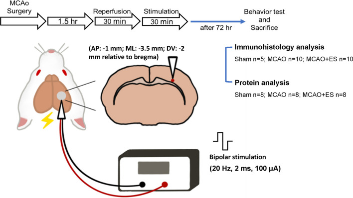
FIGURE 1.
Schematic diagram of the primary somatosensory (S1) cortical electrical stimulation (ES) experimental design. Rats sustained 90 min of transient middle cerebral artery occlusion (tMCAO) or sham operation, and electrodes were implanted to the S1 cortical area. Rats were randomly allocated to three groups and labeled as Sham (sham-operated), MCAO (tMCAO without treatment), and MCAO + ES (tMCAO with ES). A pair of electrodes was stereotactically inserted into the S1 cortex (1 mm posterior to bregma, 3.5 mm lateral to midline, and 2 mm depth) in 30 min after reperfusion. The ES-treated group received 30 min of biphasic direct current ES (frequency, 20 Hz; pulse length, 2 ms; stimulating amplitude, 100 μA; biphasic pulse) after electrode implantation. Functional outcomes were evaluated 3 days after tMCAO, and animals were sacrificed for further studies.

