FIGURE 3.
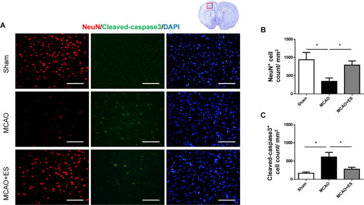
Electrical stimulation treatment inhibits neuronal cell death of the brain in the MCAO animal model. (A) Representative pictures of immunofluorescence stains for NeuN (red fluorescence), cleaved caspase-3 (green fluorescence), and DAPI (blue fluorescence). The red blocked box in the top-right panel represents views in the penumbral region. (B) Quantitative analysis for NeuN immunoreactive cells. The ES group shows significantly higher neurons survival than the control group. (C) Quantitative analysis shows a significantly reduced number of immunoreactive cells for cleaved caspase-3 in the ES group, demonstrating the antiapoptotic effect of ES (Sham n = 5; MCAO n = 10; and MCAO + ES n = 10; scale bar = 50 μm). *p < 0.05.

