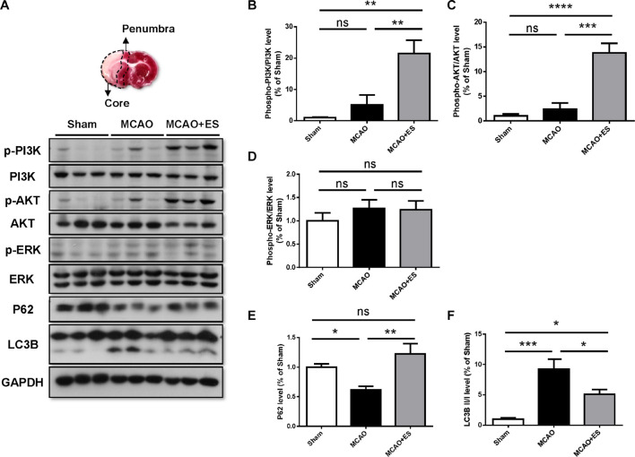
FIGURE 7.
Cortical ES treatment activated the PI3K/Akt pathway and suppressed autophagy to protect the brain from ischemia/reperfusion injury after tMCAO. Western blotting for PI3K, Akt, extracellular signal-regulated kinase (ERK), P62, and LC3B. (A) Representative pictures of Western blot. The upper panel represents the 2,3,5-triphenyltetrazolium chloride (TTC)-stained view of demarking core and penumbral region. (B,C) Phosphorylation of PI3K and Akt were significantly upregulated in ES groups. (D) There was no significant change in the phosphorylation of ERK. (E,F) In the ES group, there were increased P62 and reduced LC3B, indicating suppression of autophagy by ES (n = 5 for each group). *p < 0.05; **p < 0.01; ***p < 0.001; and ****p < 0.0001.

