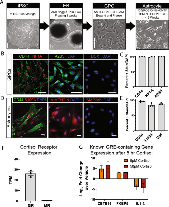
Fig. 1. iPSC-derived astrocytes differentiated in serum-free conditions express appropriate glial markers and respond to cortisol in vitro.
a Schematic of serum-free astrocyte differentiation from iPSCs via floating embryoid body (EB) and glial progenitor cell (GPC) intermediate stages. Representative bright field images depict major steps. Scale bars 100 µm. b Representative fluorescent images of immunostained GPCs expressing early glial fate markers CD44 (left-green), NF1A (left-red), and A2B5 (middle-green) and negative stain for early neuronal marker DCX (right-red); scale bars 50 µm. c Quantification of immunostain showing percent of GPCs expressing CD44, NF1A, and A2B5 out of DAPI identified total cells. Bars depict mean ± SEM; n = 3 individuals. d Representative fluorescent images of immunostained astrocytes expressing CD44 (left-green), S100β (left-red), and Vimentin (middle-red) and negative stain for neuronal marker Map2ab (red-right); scale bars 50 µm. e Quantification of immunostain showing percent of astrocytes expressing CD44, S100β, and Vimentin out of DAPI identified total cells. Bars depict mean ± SEM; n = 3 individuals. f Expression of cortisol receptors Glucocorticoid Receptor (GR) and Mineralocorticoid Receptor (MR) measured via whole transcriptome sequencing under baseline conditions. Bars show mean TPM ± SEM; n = 3 individuals. g Expression of known Glucocorticoid Response Element (GRE)-containing genes, ZBTB16, FKBP5, and L-1β, following 5 h of Cortisol or Vehicle treatment at 5 µM or 50 µM concentration. RT-qPCR values expressed as log2fold change over own vehicle control; bars depict mean ± SEM; n = 6 with 3 replicates in 2 individuals.

