
Figure 1:
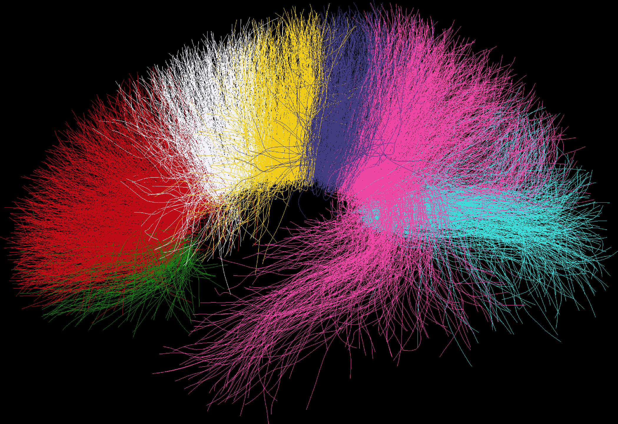
White matter (WM) fibers passing through corpus callosum (CC) subregions; green = CC1 (rostrum), red = CC2 (genu), white and yellow = CC3 + CC4 (anterior half of the body), purple and pink = CC5 + CC6 (posterior half of the body), turquoise = CC7 (splenium). WM tracts consists of grouped fiber clusters. Predefined subregions of the CC (CC1-CC7) were selected and WM fibers passing through each subregions are depicted in different colors.