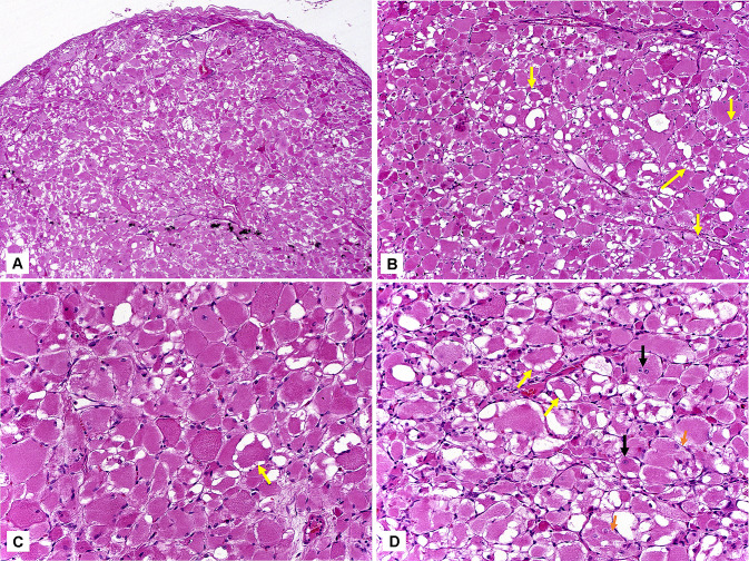
Fig. 2.
A Microscopically, at low magnification, the lesion appeared as a well-circumscribed and partially encapsulated nodule, with irregular margins (hematoxylin-eosin, 50x). B Large polygonal cells exhibited abundant, eosinophilic, and granular cytoplasm, which often presented complete or partial vacuolization; so-called “spider cells” (yellow arrows) (hematoxylin-eosin, 100x). C Large tumor cells with well-defined borders, and central or eccentric nuclei. Cytoplasmic vacuolization is also observed (“spider cells”—yellow arrows) (hematoxylin-eosin, 200x). D Most cells exhibiting granular cytoplasm, with complete or partial vacuolization (“spider cells”—yellow arrows). Prominent nucleoli (black arrows) and cross-striations (orange arrows) were also observed (hematoxylin-eosin, 200x)

