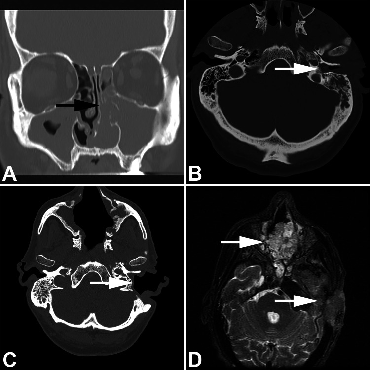
Fig. 1.
Imaging studies showing a coronal computed tomography with nasal cavity (black arrow) and maxillary sinus opacification by inverted papilloma; b middle ear (white arrow) and temporal bone opacified by papilloma; c post-surgical appearance of the ear (white arrow) and sinonasal tract; d malignant transformation identified in both the sinonasal tract (upper white arrow) and mastoid-temporal bone (lower white arrow)

