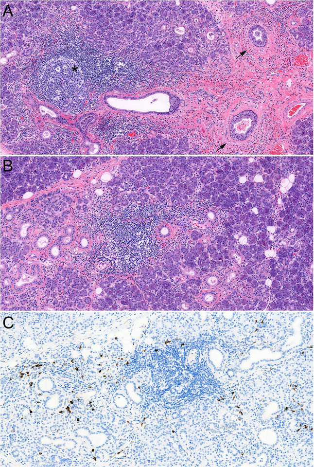
Fig. 3.

Histopathology of chronic sclerosing sialadenitis (CSS) consistent with probable/possible IgG4-related disease (comparison case, see text). There is a relatively cellular fibroinflammatory process with formation of concentric fibrosis around ducts (a, arrows) and lymphoplasmacytic inflammation with formation of germinal centers (a, asterisk). a Prominent eosinophilic infiltrate is seen in this case. b shows an acinar area with chronic inflammation and mild acinar atrophy and scattered eosinophils. c shows the same area with numerous IgG4-positive plasma cells
