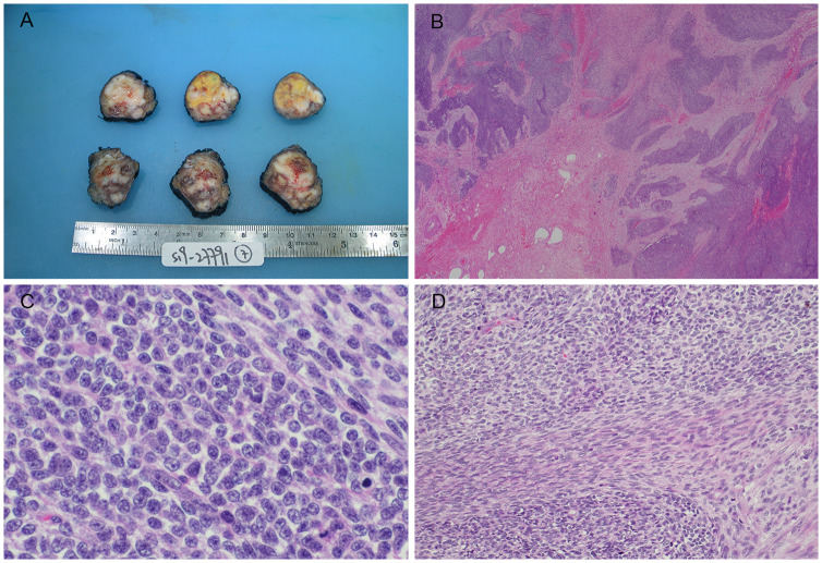
Fig. 1.
Pathological features of tumour. (a) Gross examination of tumour showing a tan-coloured to whitish solid nodular appearance with patchy hemorrhages and necrosis. (b) Microscopy revealed a lobular architecture with variably cellular fibrous stroma mimicking desmoplastic small round cell tumour. (c) Tumour featuring admixture of primitive small round cells and spindle cells, (d) some of the latter were associated with intercellular collagen

