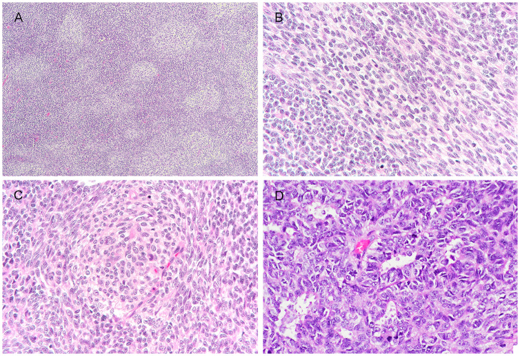
Fig. 2.
Representative images of unusual histologic features. (a) Focal “biphasic” appearance imparted by alternating pale-staining zones and small blue primitive round cell areas; The pale staining zones comprised both (b) fascicles of spindle cells with increased amount of pale staining cytoplasm and (c) small pale nodules, with focal whirling by pale spindle cells at periphery; (d) Focal rosette/ gland-like structures

