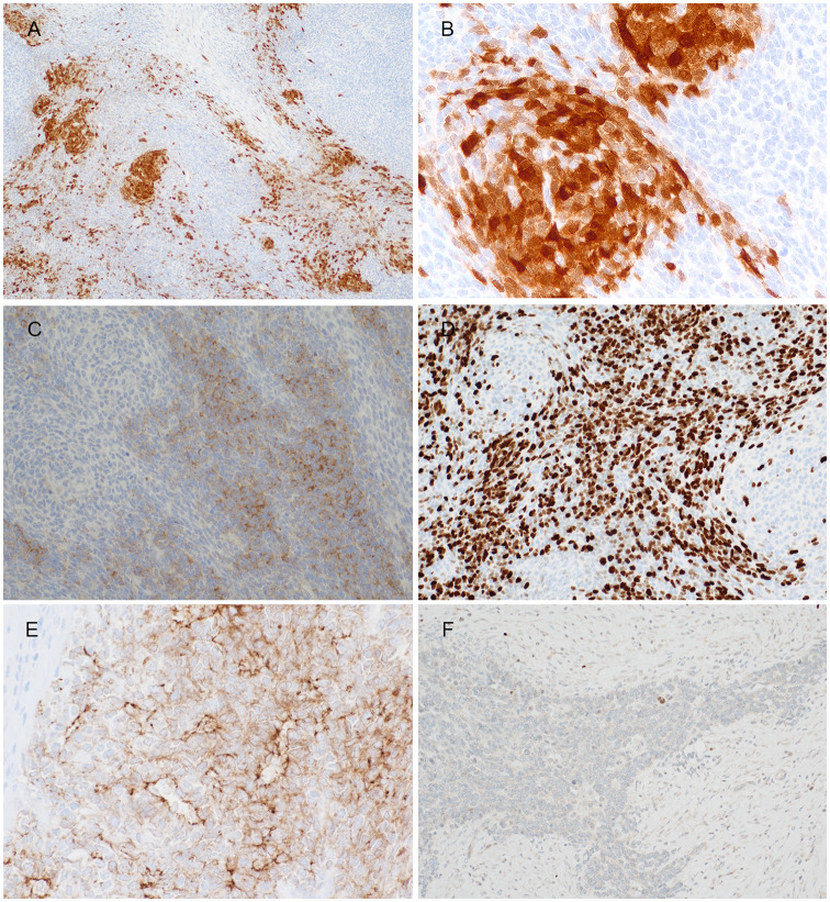
Fig. 3.
Representative images of immunohistochemistry results. S100 showed (a) patchy positivity as well as (b) focally highlighted the pale nodules. The primitive small round cells were positive to (c) synaptophysin with (d) high Ki67 index, whilst the opposite staining pattern was seen in the pale zone. (e) Claudin-4 highlighted the focal rosette/ gland-like structures, while (f) myogenin showed rare cells at the periphery of tumour lobules, probably representing entrapped skeletal muscle cells

