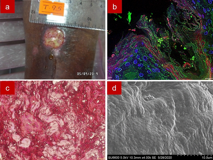
Figure 8.
Sample 27. Image of a wet ulcer of 8 weeks duration in the lower limb with green colour pus discharge (a); Fluorescence in situ hybridization showing bacteria in red with the CY3 tagged Eu bacterial probe, extracellular polymeric substances in green with Concavalin A conjugated Alexa Fluor 488 and tissue nuclei in blue with DAPI (b); Gram staining showing gram-negative rods (c); Scanning electron microscopy showing bacteria in embedded in smooth extracellular polymeric substance (d).

