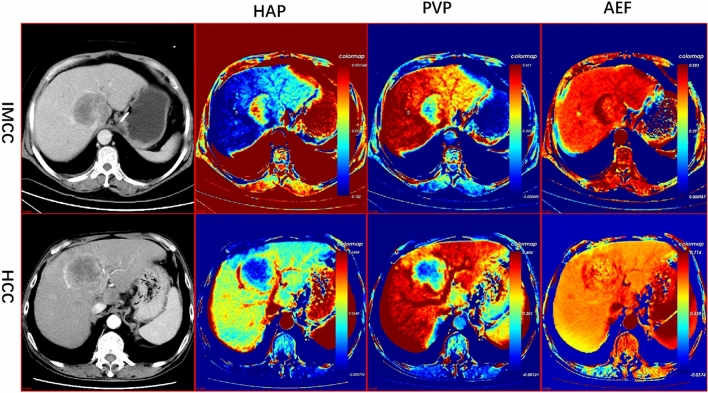
Figure 2.
Traditional enhancement image and pharmacokinetic images of IMCC and HCC. For the patients with IMCC, the HAP image showed high perfusion in the margin and relatively low perfusion in the center. PVP images showed hyperperfusion from the peripheral to the central part of the tumor. For patients with HCC, the HAP image showed high perfusion in the rim, while the PVP image showed homogeneous low perfusion in the complete lesion. The AEF images both showed heterogeneous high perfusion for two tumor lesions. HAP Hepatic arterial supply perfusion, PVP Portal venous supply perfusion, AEF Arterial enhancement fraction, IMCC Intrahepatic mass-forming cholangiocarcinoma, HCC Hepatocellular Carcinoma.

