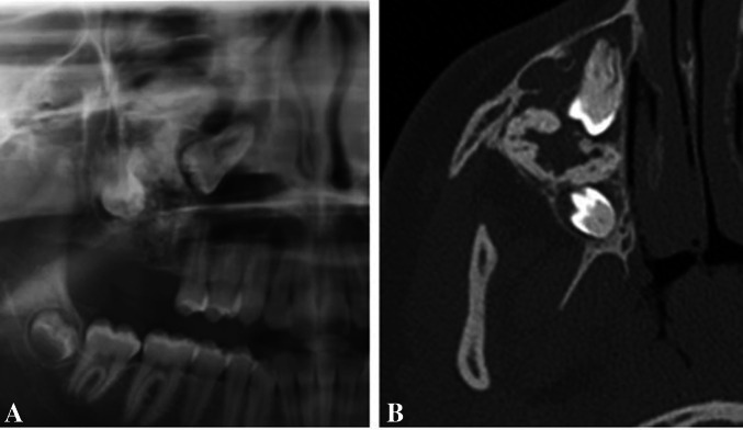
Fig. 2.
A Cropped panoramic view of an AFO showing a large, mixed radiolucent-radio-opaque lesion in the right maxilla. Margins are difficult to be defined. The first and second molars are involved and displaced. B Axial plane cone-beam computed tomography image highlights the extent of the lesion and its margins. A few bony septae denote the lesion a multilocular appearance

