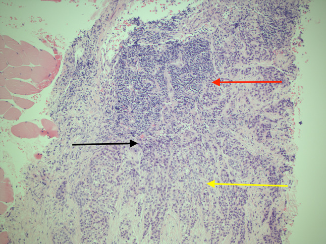
Fig. 5.

Histopathologic slides showing the lymph node core biopsy. H&E stain, ×100 magnification. Demonstrates sheets of polyhedral epithelial cells adjacent to lymphoid tissue of the lymph node. Red arrow shows lymphoid tissue. Yellow arrow shows clusters of clear cells with pleomorphism and hyperchromatic nuclei. Black arrow shows epithelial cells with pleomorphism and hyperchromatic nuclei adjacent to the lymphoid tissue. Note also the small blood vessel within the area with malignant cells surrounding the vessel
