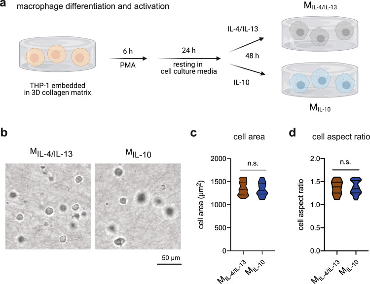
Fig. 1. Macrophage differentiation and activation in 3D collagen matrices.
a Schematic workflow for macrophage differentiation and activation towards MIL-4/IL-13 and MIL-10 in 3D collagen matrix. b Representative images of MIL-4/IL-13 and MIL-10 were gathered using bright-field microscopy. A quantitative morphological analysis, namely c cell area and d cell aspect ratio, was conducted using an image analysis toolbox. Data are presented as a violin plot. At least 100 cells from three independent experiments were analyzed.

