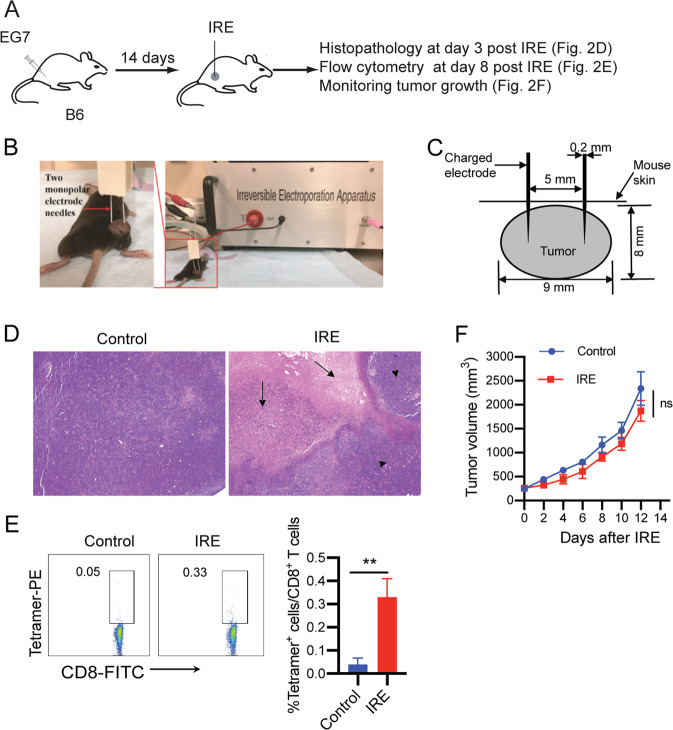
Fig. 2. IRE ablation induces tumor cell apoptosis but weak OVA-specific CD8+ T cell responses and is ineffective in inhibiting tumor growth.
A Diagram illustrating the design of the IRE ablation experiment. B Experimental setup for IRE treatment. C Schematic diagram showing the placement of the IRE device electrode in a tumor (8–9 mm in diameter) during IRE ablation. D Representative H&E staining of tumor tissue sections collected at 3 days post IRE ablation. Arrows indicate areas of massive apoptosis in IRE-treated tumors. Arrowheads indicate the surrounding tumor tissues. E Blood cells collected from the tail vein of IRE-treated or naïve control mice were stained with OVA-specific PE-Tetramer and a FITC-conjugated anti-CD8 antibody and analyzed by flow cytometry. OVA-specific CD8+ T cells were defined as CD8 and tetramer double-positive cells. The value in each panel represents the percentage of OVA-specific CD8+ T cells in the total CD8+ T cell population. **P < 0.01 by two-tailed Student t test. F Tumor-bearing mice were monitored for tumor growth post IRE ablation. ns, not significant by two-way ANOVA with Tukey’s test. Flow cytometry or tumor growth plots representing one of two independent experiments are presented as the mean ± SEM (n = 5/group)

