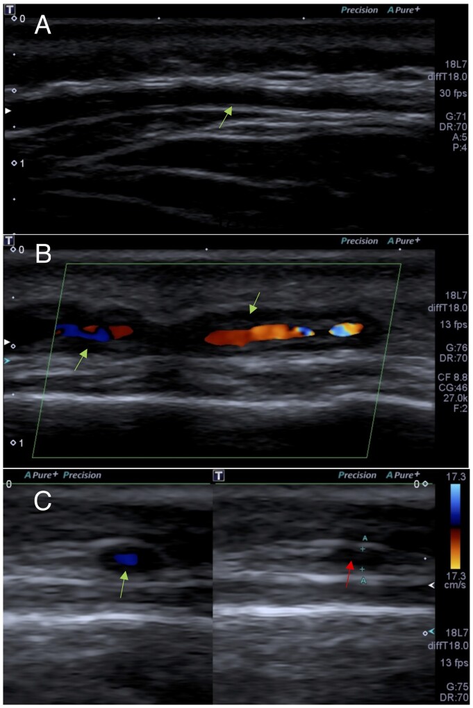
Fig. 3.
Colour Doppler ultrasonography of the temporal arteries
(A) Longitudinal view of a normal intima–media complex of the left frontal branch of the temporal artery (arrow). (B) Longitudinal view of a hypoechoic thickening of the intima–media (halo sign) of the left parietal branch of the temporal artery (arrow). (C) Transverse view of a halo sign using colour Doppler (green arrow) and positive compression sign in that location (red arrow).

