Figure 1.

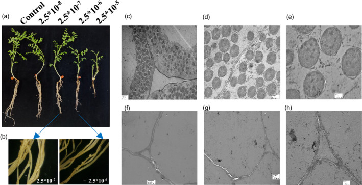
Effect of exogenous cytokinin treatment in chickpea seedlings (a) Phenotypic appearance of 30‐day‐old chickpea seedlings under different concentration of 6‐BA [2.5 × 10⁻⁸ to 2.5 × 10⁻⁵M]. (b) Stereo zoom microscopic images are showing pseudo‐nodules observed in 2.5 × 10⁻⁷M and 2.5 × 10⁻⁶M (scale bar = 50 pixel) without rhizobia infection. Control: no treatment with cytokinin and rhizobia; 6‐BA concentration: 2.5 × 10⁻⁸ to 2.5 × 10⁻⁵M. (c‐e) TEM images of true chickpea containing bacteriods with M. ciceri infection. [Scale bar = 2 µm, 500nm and 100 nm, respectively, and magnification = 1000×, 2500× and 6000×, respectively]. (F‐H) TEM images of chickpea pseudo‐nodules in cytokinin concentration of 2.5 × 10⁻⁷M in the absence of M. ciceri infection. [Scale bar = 2 µm 2 µm and 500nm, respectively and magnification = 1000×, 1000× and 2500×, respectively]. Experiments were carried in three biological replicates in ten chickpea seedlings.
