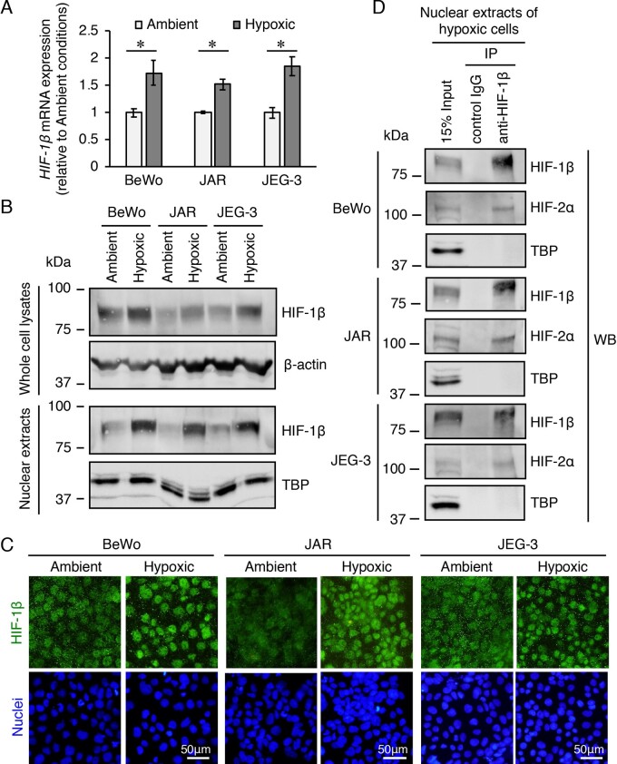
Figure 1.
mRNA and protein expression levels of hypoxia-inducible factor (HIF)-1β and the interactions between HIF-1β and HIF-2α in three trophoblast-derived choriocarcinoma cell lines exposed to hypoxia. BeWo, JAR and JEG-3 cells were incubated under ambient (atmosphere with 5% CO2) or hypoxic (2% O2, 5% CO2 and 93% N2) conditions for 24 h. (A) The mRNA expression levels of HIF-1β were measured by quantitative reverse transcription-polymerase chain reaction using β-actin mRNA as a reference. Results are expressed as the fold change relative to the cells under ambient conditions. All values are represented as the mean ± SD (n=3). Asterisks indicate the significant difference (P<0.05). (B) The expression levels of HIF-1β in the whole cell lysates and nuclear extracts of the three cell lines were assessed by western blotting analysis. β-Actin and the nuclear protein, TATA binding protein, were used as the loading controls. (C) Cellular localization of HIF-1β in the three cell lines was determined by immunofluorescence staining. (D) Immunoprecipitation of HIF-2α protein using an antibody against HIF-1β in nuclear extracts of the three hypoxic cell lines. The immunoprecipitates and nuclear extracts (input) were subjected to western blotting analysis. Uncropped images of the western blots are presented in Supplementary Fig. S4.

