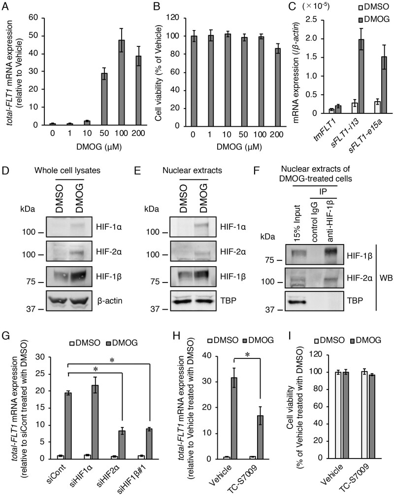
Figure 3.
Role of HIFs in the upregulation of the FLT1 gene under chemically mimicked hypoxic conditions using the HIF-α stabilizer dimethyloxalylglycine (DMOG) in BeWo cells. (A) Effect of various doses of DMOG on the mRNA expression levels of all FLT1 transcript variants (total-FLT1). The cells were treated with various doses of DMOG for 24 h. Results are expressed as the fold change relative to the vehicle (dimethyl sulfoxide (DMSO))-treated cells. (B) Cell viability was assessed to determine the potential toxicity of DMOG. Results are expressed as the percentage relative to the vehicle-treated cells. (C) The mRNA expression levels of the three FLT1 splice variants in the presence of 100 µM DMOG or 0.1% DMSO (vehicle control). (D, E) Western blotting analysis of the expression levels of HIF proteins in the whole cell lysates and nuclear extracts of the vehicle- or 100 µM DMOG-treated cells. β-Actin and TATA binding protein were used as the loading controls. (F) Immunoprecipitation of HIF-2α protein using an antibody against HIF-1β in the nuclear extracts of the 100 µM DMOG-treated cells. The immunoprecipitates and nuclear extracts (input) were subjected to western blotting analysis. Uncropped images of the western blots are presented in Supplementary Fig. S4. (G) Effects of the gene silencing of each HIF on DMOG-induced upregulation of the FLT1 gene. Cells were transfected with the indicated siRNAs targeting HIFs for 72 h prior to treatment with either DMSO or DMOG for 24 h. Results are expressed as the fold change relative to the siCont-transfected cells in the presence of the vehicle. (H) Effect of the HIF-2α inhibitor, TC-S7009, on DMOG-induced upregulation of the FLT1 gene. The cells were cultured for 24 h in the presence of 100 µM DMOG or the vehicle in the presence of 0.1% DMSO (vehicle control) or 30 µM TC-S7009. Results are expressed as the fold change relative to the DMSO-treated cells in the presence of the vehicle. (I) Cell viability was assessed to determine the potential toxicity of TC-S7009. Results are expressed as the percentage relative to the DMSO-treated cells in the presence of the vehicle. All values are represented as the mean ± SD (n=3). Asterisks indicate the significant difference (P<0.05).

