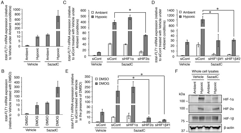
Figure 4.
Both HIF-2α and HIF-1β regulate the further increase in the expression levels of the FLT1 gene induced by hypoxia or DMOG in the BeWo cells treated with a DNA methyltransferase inhibitor 5′-aza-2′-deoxycytidine (5azadC). (A, B) The cells were incubated in the presence of 0.1% DMSO (vehicle control) or 10 μM 5azadC and the culture media were changed daily. After 3 days of culture, the culture media were changed with fresh growth media without 5azadC. The cells were treated under hypoxia or with DMOG for 24 h and then the mRNA expression levels of all FLT1 transcript variants (total-FLT1) were measured by quantitative reverse transcription-polymerase chain reaction using β-actin mRNA as a reference. Results are expressed as the fold change relative to the control vehicle-treated cells under ambient conditions and in the presence of DMSO. (C–E) The cells that underwent demethylation and siRNA transfection simultaneously for 72 h were subjected to hypoxic or chemically mimicked hypoxic conditions for 24 h, after which the mRNA expression levels of total-FLT1 were determined. Results are expressed as the fold change relative to the siCont-transfected cells treated with the vehicle under ambient conditions and in the presence of DMSO. All values represent the mean ± SD (n=3). Asterisks indicate significant differences (P<0.05). (F) Western blotting analysis of the expression levels of HIF proteins in the whole cell lysates of the BeWo cells, with or without 5azadC treatment, under ambient and hypoxic conditions. β-Actin was used as the loading control. Uncropped images of the western blots are presented in Supplementary Fig. S4.

