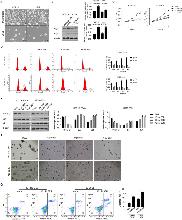
Figure 1.
Berberine inhibited malignancies and induced apoptosis in HCT116 and HT29 CSCs. (A) Parental HCT116 and HT29 cells were cultured in serum-free medium for 14 days, and spheres were obtained. (B) CD44 and CD133 protein levels were detected by performing the Western blot in total protein. (C) By being cocultured with 10, 20, or 40 μM of Berberine for 1 to 5 days, cell viability was measured by performing CCK-8 assay. p < 0.05, vs. mock group. By being cocultured with 10, 20, or 40 μM of Berberine for 24 h, cell cycle distribution was analyzed by performing flow cytometry assay (D) and p21, p27, and Cyclin D1 protein levels were measured by performing Western blot (E). p < 0.05, vs. mock group. (F) Tumor formation ability of CSCs was measured by being cocultured with 10, 20, or 40 μM of Berberine for 14 days. (G) By being cocultured with 10, 20, or 40 μM of Berberine for 24 h, cell apoptosis was measured by performing Annexin V-FITC/PI double staining. *P < 0.05, vs. mock group.

