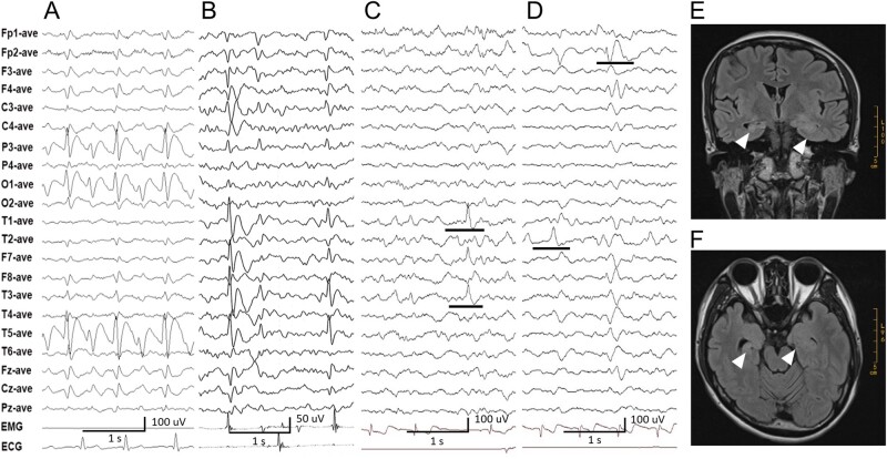
Figure 4.
Representative EEG recordings and MRI from patients with UNC13B variants. (A) Interictal EEG in Case 1 showed left occipital slow spike waves. (B) Interictal EEG in Case 3 showed left central-temporal slow spike waves. (C and D) Interictal EEG in Case 8 showed spikes or sharp waves in the left or right temporal lobe, or of slow spike waves in the right frontal lobe. (E and F) Coronal and axial T2-FLAIR MRI of Case 8 revealed structural asymmetry in the hippocampus; the right hippocampus was smaller than the left one, with a slightly higher signal. The boundary between the left hippocampus and surrounding tissues was indistinct.

