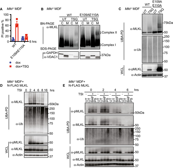
Figure 5. MLKL ubiquitylation does not lead to cell death, but correlates with the turnover of activated MLKL.

- WT and E109AE110A mutant MLKL were inducibly expressed in Mlkl −/− MDFs by doxycycline, and cells were untreated (UT) or treated with TSQ for 6 h (please note that the same WT control was used in Fig EV3A). Cell death was measured by PI staining based on flow cytometry. Data are plotted as mean ± SEM of three independent experiments.
- Cytosol (C) and crude membrane (M) cellular fractions from (A) were analysed by Western blot from BN‐PAGE or SDS–PAGE using antibodies as indicated. The same WT control was used in Fig 4A. Representative of three independent experiments.
- Cell lysates from (A) were subjected to UBA pulldown and analysed as described above.
- N‐FLAG MLKL were inducibly expressed in Mlkl −/− MDFs by doxycycline overnight, and cells were treated with TSI for indicated time, followed by UBA pulldown. Representative of three independent experiments.
- N‐FLAG MLKL were inducibly expressed in Mlkl −/− MDFs by doxycycline for 16 h. Cells were stimulated ± TSI for 3 h after withdrawal of doxycycline. Then, TSI medium was removed and replaced to medium containing inhibitors Bafilomycin A1 (BAF), PS341 (PS) or left untreated (UT). IDN‐6556 was added to all conditions to block apoptosis. Cells were collected 0, 2, 4 and 6 h after medium replacement, followed by UBA pulldown. Representative of three independent experiments.
Data information: Wild‐type (WT). TSI and TSQ are used as necroptotic stimuli.
Source data are available online for this figure.
