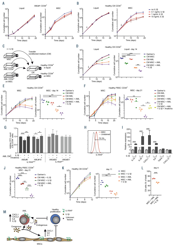
Figure 5.
The IL1-IL1RAP signaling pathway affects normal hematopoiesis but not acute myeloid leukemia cell growth in the context of human mesenchymal stromal cells. (A) Growth curve of acute myeloid leukemia (AML) patient 1 (AML#1) CD34+ cells in liquid culture (left) and on a stromal layer of mesenchymal stromal cells (MSC) (right) ±IL1b in different concentrations. The arrow indicates from whereon IL1b was added. (B) Growth curve of CB CD34+ cells in liquid culture (left) and on a stromal layer of MSC (right) ±IL1b in different concentrations. The arrow indicates from whereon IL1b was added. (C) Schematic overview of experimental setup of co-cultures and conditioned medium (CM) transferring. (D and E) Growth curve (left) and cumulative cell number on day 14 (right) of CB CD34+cells in liquid (D) and on a stromal layer of MSC (E) with the addition of CM from a MSC culture (CM MSC), AML#1 CD34+ liquid culture (CM AML), AML#1 CD34+ MSC co-culture (CM MSC + AML), and AML#1 CD34+ MSC co-culture with the addition of 10 ng/mL IL1b (CM MSC + AML + IL1b). CM was added at day 4 (arrow) and every following demi-population. (F) Growth curve (left) and cumulative cell number on day 21 (right) of CD34+ peripheral blood stem cells (PBSC) grown on MSC with the CM from AML#18 (co-)cultures. Treatment conditions were similar to the once described in the legend of Figure 5D and E, adding one condition including AML CD34+ co-culture with the addition of 10 ng/mL IL1b and 500 ng/mL Anakinra (CM MSC + AML + IL1b + Anakinra). CM was added at day 0 and at every following demi-population. (G) Colony-forming cell (CFC) assay of cord blood (CB) CD34+ treated with CM of IL1RAPhigh AML (AML#9 and AML#19) and IL1RAPlow AML (AML#20 and AML#21), which were cultured for 7 days on a MSC stromal-layer in the presence or absence of IL1b and Anakinra, before CM was harvested. Data of two biological duplicates are shown relative to the untreated condition. (H) Interleukin-1 receptor accessory protein (IL1RAP) expression on MSC measured by flow cytometry. (I) Quantitative real-time polymerase chain reaction of MSC stimulated with and without IL1b. Statistical analysis was performed using a Student’s t-test. (J) Cumulative cell number on day 21 of CD34+ PBSC grown on MSC (experimental setup identical to panel F) including conditions AML + IL1b and MSC + IL1b. Gartner’s and CM MSC + AML + IL1b (identical to panel F) has been added for direct comparison. (K) Growth curve (left) and cumulative cell number on day 11 (right) of CB CD34+ cells in triple co-culture with MSC and AML#16 CD34+ cells ±IL1b (L) Percentage of CB cells in triple co-culture with MSC and AML#22 ±IL1b at day 4. (M) Schematic model how AML cells might impact on normal hematopoiesis in the bone marrow niche, in part via the IL1-IL1RAP axis. Statistical analysis in all panels was performed using a Student’s t-test.* P<0.05; **P<0.01; ***P<0.001.

