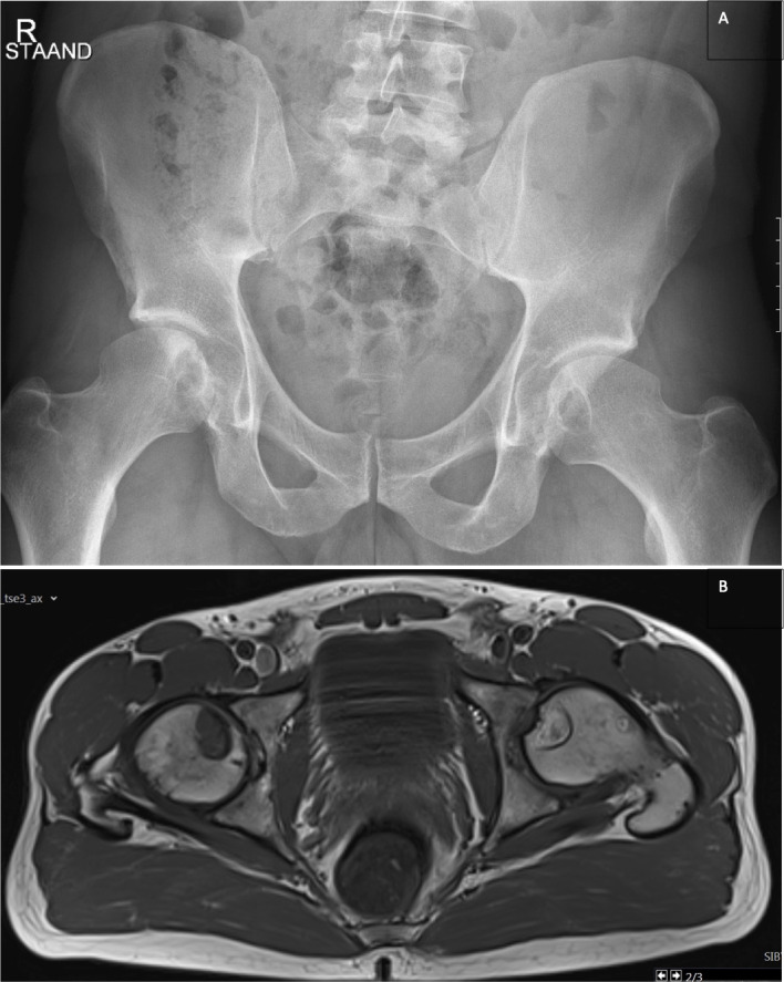
Figure 1.

(A) Anteroposterior radiographs of the right (and left (hips. Bilateral subchondral cysts in the inferior half of the femoral heads. (B) T1W axial image showing isointense aspect of the cyst on the right side with perilesional discrete isolated bone marrow oedema and inhomogeneous hyperintense aspect of the cyst on the left-side suspect for a mucoid (chronic) component. Both 'lesions' are characterised by intralesional pencil-fine (osseous lamellar) septations and a sharp limited sclerotic peripheral contour delineation. note also the globally preserved sphericity of the femoral heads bilateral with no apparent collapse.
