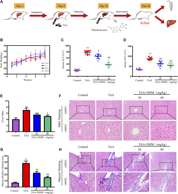
FIGURE 1.
DHM ameliorated TAA-induced hepatic injury in mice. The experimental design (A); Body weight (B) and serum levels of ALT (C) and AST (D) (n = 8); liver index (E), H&E staining (F); Masson relative stained area (G) and Masson’ strichrome staining (H); Data was expressed as the mean ± SD. The differences between groups were analyzed by two tailed T-test or one-way analysis of variance (ANOVA). **p < 0.01, *p < 0.05 versus control group; ##p < 0.01, #p < 0.05 versus TAA group.

