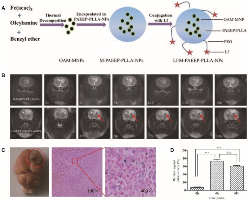
Figure 4.
Lf-M-PAEEP-PLLA-NPs were used as glioblastoma-targeted MRI contrast agents [33]. (A) Schematic illustration of Lf-M-PAEEP-PLLA-NP preparation. (B) In vivo MRI of C6-bearing rats. (C) Prussian blue staining assays of c6-bearing rats after Lf-M-PAEEP-PLLA-NP injection. (D) Quantification of the signal enhancement in the tumour area at different time points. Reproduced with permission [33]. Copyright 2016, Springer Nature. Lf, lactoferrin; MRI, magnetic resonance imaging; NPs, nanoparticles; PAEEEP-PLLA, amphiphilic poly(aminoethyl ethylene phosphate)/poly(L-lactide)

