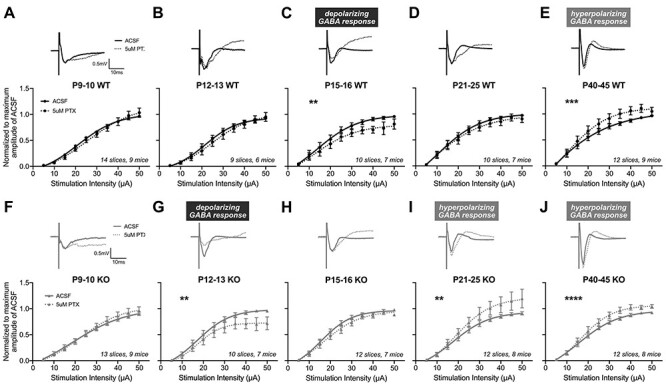
Figure 2 .

The auditory cortex of Fmr1 KO mice exhibits an accelerated age-dependent shift in their sensitivity to PTX. I–O curves from MEA field recordings were analyzed following 5 μM PTX. (A–E) Top: representative traces from Fmr1 WT mice at 40 μA stimulation. Bottom: I–O curves before and after PTX, normalized to the maximum amplitude in ACSF and fit with sigmoidal curves. (C) P15–16 Fmr1 WT mice exhibit decreased fEPSP amplitudes following PTX, suggestive of the presence and subsequent blockade of depolarizing GABA by PTX. (E) Fmr1 WT exhibit significantly increased fEPSP amplitudes to PTX at P40–45, indicating PTX’s blockade of inhibitory hyperpolarizing GABA. (F–J) Representative traces and I–O curves from Fmr1 KO mice. (G) Fmr1 KO mice exhibit significantly decreased responses to PTX at P12–13 but not at (H) P15–16. (I–J) At both P21–25 and P40–45, Fmr1 KO mice exhibit significantly increased amplitudes to PTX. The number of slices and mice analyzed for each age and genotype are indicated in each graph. **P < 0.01, ***P < 0.001, ****P < 0.0001.
