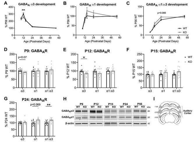
Figure 3 .

Fmr1 KO mice exhibit altered developmental regulation in the expression of GABAAR α1 and α3 subunits in the auditory cortex. (A–C) Western blot analysis of membrane-bound expression of GABAAR α3, GABAAR α1, and GABAAR α1:α3, with all timepoints normalized to P56 WT to analyze the developmental expression of the GABAAR α-subunits in the auditory cortex. (A) Developmental expression of GABAAR α3 exhibits an overall interaction between age and genotype, with significantly decreased expression for KO compared with WT at P12. (C) Developmental GABAAR α1:α3 expression exhibits an overall interaction between age and genotype. (D–G) GABAAR α-subunits expressed relative to age-matched WT littermates. (D) Fmr1 KO exhibit a trending decrease in GABAAR α3 expression at P9 (n = 11 WT, 11 KO). (E) GABAAR α3 is significantly decreased at P12 (n = 10, 11). (F) No differences in expression observed at P15 (n = 10, 10). (G) P24 Fmr1 KO exhibit a trending increase in GABAAR α1, with significantly increased GABAAR α1:α3 expression (n = 12, 11). No differences were observed at P56 (n = 18, 15), data not shown. (H) Representative western blots of GABAAR α3 and GABAAR α1 with β-actin used as loading controls. (I) Schematic of approximate auditory cortex dissections as described in Materials and Methods. *P < 0.05, **P < 0.01.
