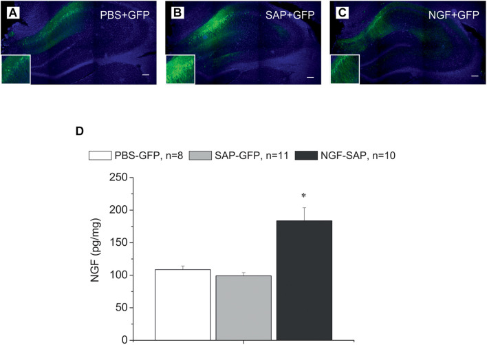
FIGURE 1.
Immunostaining of hippocampal slices with antibody against GFP and DAPI staining in the hippocampus control rats (A) and rats with cholinergic deficit (B,C), which were transduced with AAV carrying either GFP alone (A,B) or NGF-IRES-GFP (C). The images of GFP staining (green) are superimposed over images of the same sections stained with DAPI (blue). (D) Shows NGF protein concentration determined by ELISA in the hippocampi of control (PBS-GFP, n = 8), 192IgG-saporin (SAP-GFP, n = 11), and NGF overexpressed rats (SAP-NGF, n = 10). Each point represents the mean ± SEM. ∗Significant differences compared to PBS-GFP and SAP-GFP groups, p < 0.05; bar, 200 μm.

