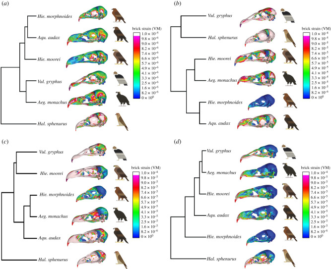
Figure 2.
VM strain contour plots of the crania in the intrinsic, dorsoventral, lateral shake and pullback load cases in lateral view and UPGMAs performed on PC scores of VM strains collected at specific landmarks for each of the considered load cases: (a) intrinsic, (b) dorsoventral, (c) lateral shake and (d) pullback. Models are not to scale. Bird art by Scott Partridge. (Online version in colour.)

