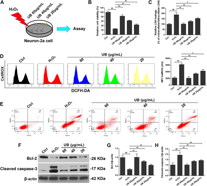
FIGURE 1.
(A) The schematic diagram of the cell experimental design for H2O2-induced Inflammatory injury model and UB treatment. The protective effect of UB on neuron-2a cells against H2O2-induced viability (B) and cytotoxicity (C). (D) UB inhibited ROS accumulation in neuron-2a cells, as determined by flow cytometry. (E) The anti-apoptotic effect of UB against H2O2-induced neurotoxicity in neuron-2a. (F) The expression of cleaved caspase-3 and Bcl-2 was detected by western blotting. Quantification analysis of cleaved caspase-3 (G) and Bcl-2 (H). Data were normalized to β-actin protein expression. All results are described as means ± SD (n = 3 per group). # p < 0.05 and ## p < 0.01 vs the control group; * p < 0.05 and ** p < 0.01 vs the H2O2-treated group.

