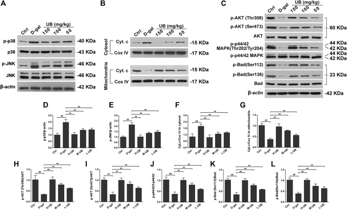
FIGURE 6.
(A) Western blots and densitometric analysis of the phosphorylation of stress-activated protein kinase (SAPK)/c-Jun NH 2-terminal kinase (JNK) and p38 MAPK in the brains of aging mice. (B) Western blots and densitometric analysis of cytosolic and mitochondrial Cyt C and COX IV in all treated groups. (C) Western blots and densitometric analysis of the phosphorylation of Akt, p44/42 MAPK and Bad in the brains of D-gal-treated aging mice. Quantification analysis of p-p38 (D), p-JNK (E), Cyt.c/COX IV in cytosolic (F) and mitochondrial (G), p-Akt (Thr308)/Akt (H), p-Akt (Ser473)/Akt (I), p-p44/42/t-p44/42 (J), p-Bad (Ser112)/Bad (K) and p-Bad (Ser136)/Bad (L). Data were normalized to β-actin protein expression (n = 6 per group). All results are described as means ± SD. # p < 0.05 and ## p < 0.01 vs the control group; * p < 0.05 and ** p < 0.01 vs the D-gal-induced aging group.

