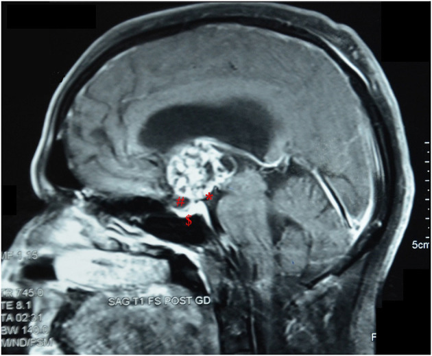
Figure 2.

Post-contrast sagittal MRI image of a patient with an intrinsic third ventricular craniopharyngioma, showing features as described by Migliore et al. (16). *, an intact third ventricular floor; #, a patent suprasellar cistern; $, absence of sellar abnormalities.
