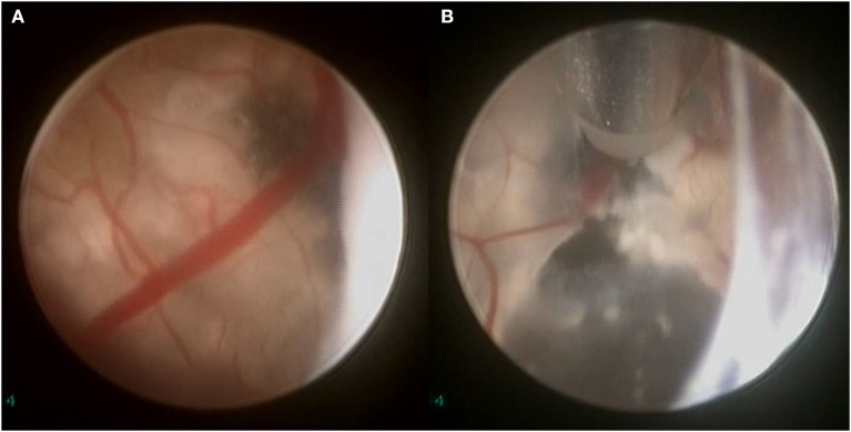
Figure 5.
Intraoperative endoscopic view of a cystic intraventricular craniopharyngioma in a 6-year-old child. (A) Craniopharyngioma cyst wall (with calcifications) visualized. (B) Using bipolar diathermy, a fenestration is made into the cyst wall for draining its contents and insertion of a ventricular catheter connected to an Ommaya reservoir (through which alpha-interferon was administered).

