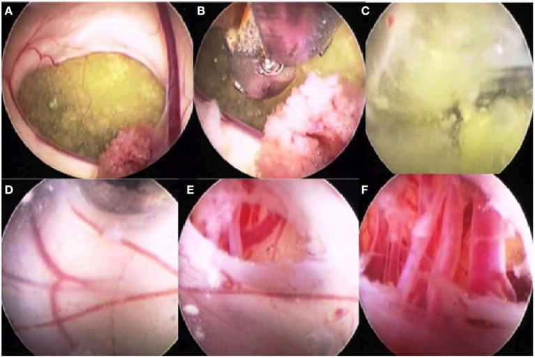
Figure 8.
Intraoperative images of a patient with a cystic intraventricular craniopharyngioma undergoing endoscopic cyst fenestration and third ventriculostomy. (A) Intra-third ventricular craniopharyngioma cyst seen through foramen of Monro. Choroid plexus is also seen. (B) The cyst is being fenestrated using an endoscope through the frontal, transcortical, transforaminal approach. (C) The machinery oil fluid of the craniopharyngioma cyst is well seen. (D) The inner wall of the craniopharyngioma cyst blended with the third ventricular ependymal floor is seen after drainage of the cyst contents. The cyst wall along with the third ventricular floor is being fenestrated. (E) The arterial blood vessels in the interpeduncular cistern are visible. (F) The endoscope tip is taken through the inferior cyst wall and the ventricular ependyma to see the blood vessels of the interpeduncular cistern.

