Fig. 5.

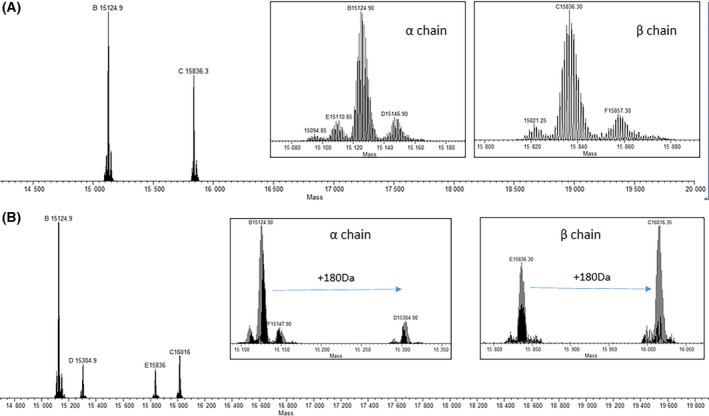
Deconvoluted ESI‐MS spectra of (A) HbS and (B) HbS + CA. The +180 m/z mass shift increase unique to HbS +CA in both the α‐chain and β‐chain indicates covalent bonding of CA to each HbS chain.

Deconvoluted ESI‐MS spectra of (A) HbS and (B) HbS + CA. The +180 m/z mass shift increase unique to HbS +CA in both the α‐chain and β‐chain indicates covalent bonding of CA to each HbS chain.