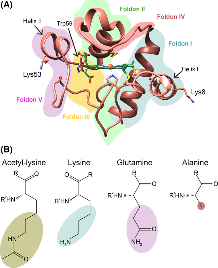
Fig. 1.

3D structure of cytochrome c and formula of selected amino acids. (A) Ribbon representation of human Cc structure (PDB, ID: 2N9I [42]), with the heme group (green color) and Lys8, Lys53, and Trp59 represented by sticks. The different foldon units are colored as follows: foldon I in blue, foldon II in green, foldon III in yellow, foldon IV in red, and foldon V in purple. (B) Chemical structure of acetyl‐lysine, lysine, glutamine, and alanine.
