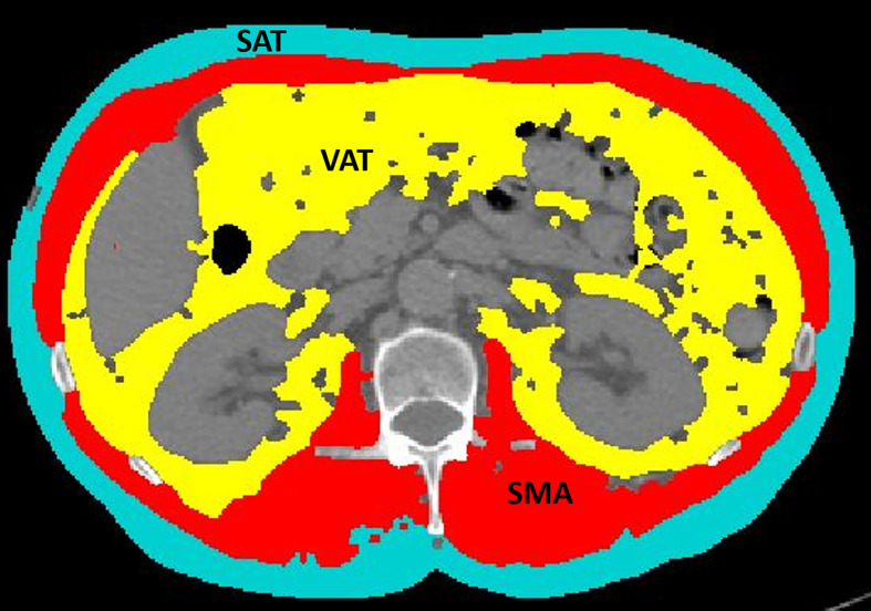
Figure 1.
An example of segmentation of skeletal muscle area (red), visceral adipose tissue (yellow), and subcutaneous adipose tissue (light blue) from an axial image at the level of L3 of a CT scan performed as routine pretreatment staging. The colored parts are quantified as areas (cm2) by the software. The excluded parts include abdominal organs, such as liver, kidneys, and bowel (dark gray), as well as bones, such as vertebral body and ribs (light gray and white).

