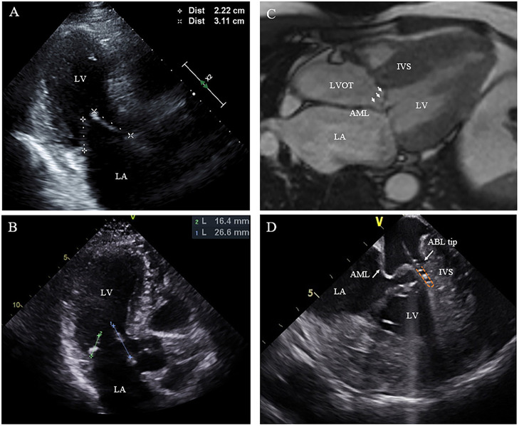
Figure 3.
Morphologic abnormalities of mitral valve contributing to outflow tract obstruction. (A,B) The length of the AML was measured by echocardiography in diastole on the apical three-chamber view. (C) Extraordinarily long anterior mitral valve leaflet on CMR. (D) On live ICE screen, the AML (white arrow) is reflexed and attached to IVS during systole and pats on the ablation catheter tip during procedure. AML, anterior mitral leaflet.

