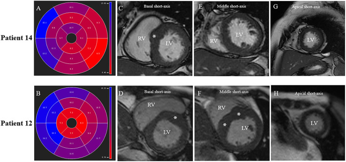
Figure 4.
Different patterns of septum hypertrophy among patients. Patient No.12: (B,D,F,H). Patient No.14: (A,C,E,G). (A,B) CMR end-diastolic images demonstrating different patterns of interventricular septum hypertrophy. The distribution of LV hypertrophy is limited (basal anteroseptal segment) in (A), while diffused (basal, mid anteroseptal, and mid inferoseptal segments) in (B). (C,D) Short-axis view presents a segmental LV hypertrophy of the basal anterior septum in both patients. (E,F) Both mid anteroseptal and inferoseptal segments are free of hypertrophy in (E), but present hypertrophied phenotype in (F). (G,H) Apical septal segment is free of hypertrophy in both patients.

