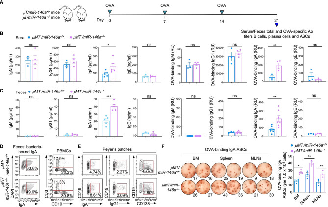
Figure 3.
Increased IgA production, IgA+ B cells and IgA-ASCs in μMT/miR-146a–/– chimeric mice. μMT/miR-146a–/– and μMT/miR-146a+/+ chimeric mice were constructed by mixed bone marrow adaptive transfer. (A) All mice were administered OVA together with CT via intragastric gavage once a week for 3 wks and sacrificed one wk after the last OVA administration for analysis of antibodies, B cells and ASCs. Total and OVA-binding IgM, IgG1, IgA and IgE titers in sera (B) and feces (C) as analyzed by ELISAs. (D) Fecal bacteria-bound IgA as analyzed by flow cytometry. (E) Circulating T (CD3+) and B (CD19+) cells, Peyer’s patched IgA+ B cells, IgG1+ B cells and CD138+ plasma cells, as analyzed by flow cytometry. (F) ELISPOT analysis of OVA-binding IgA ASCs in the bone marrow (BM), spleen and MLNs. ***p < 0.001, **p < 0.01, *p < 0.05, ns, not significant, unpaired t-test. Data in (B, C) and (F, right panel) are mean ± SEM of 3-7 μMT/miR-146a–/– or μMT/miR-146a+/+ from three independent experiments. Data in (D, E) and (F, left panel) are from one representative of three independent experiments yielding similar results.

