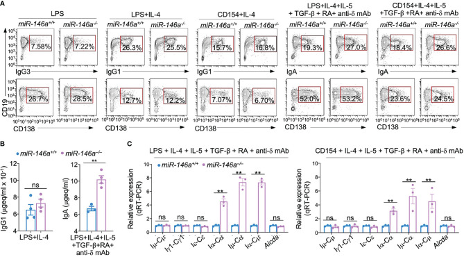
Figure 5.
miR-146a ablation results in increased germline Iα-Cα transcripts and enhanced CSR to IgA. (A) miR-146a–/– and miR-146a+/+ B cells were stimulated with LPS, LPS or CD154 plus IL-4, LPS or CD154 plus IL-4, IL-5, TGF-β, RA and anti-δ mAb, and cultured for 96 h. IgG1+, IgG3+ and IgA+ B cells and CD138+ plasma cells as analyzed by flow cytometry. Data are one representative of three independent experiments yielding similar results. (B) IgG1 and IgA concentrations in culture fluids from 146a–/– and miR-146a+/+ B cells stimulated by LPS plus IL-4 or LPS plus IL-4, IL-5, TGF-β, RA and anti-δ mAb, as measured by ELISA. (C) miR-146a–/– and miR-146a+/+ B cells were stimulated by LPS or CD154 plus IL-4, IL-5, TGF-β, anti-δ mAb and RA, and cultured for 72 h. Expression of germline Iμ-Cμ, Iγ1-Cγ1, Iα-Cα and Iϵ-Cϵ transcripts, circle Iα-Cμ and post-recombination Iμ-Cα transcripts, as well as Aicda transcripts as analyzed by qRT-PCR and normalized to β-Actin. Data are ratios to miR-146a+/+ B cells (set as 1; means ± SEM of three independent experiments). **p < 0.01, ns, not significant, paired t-test.

