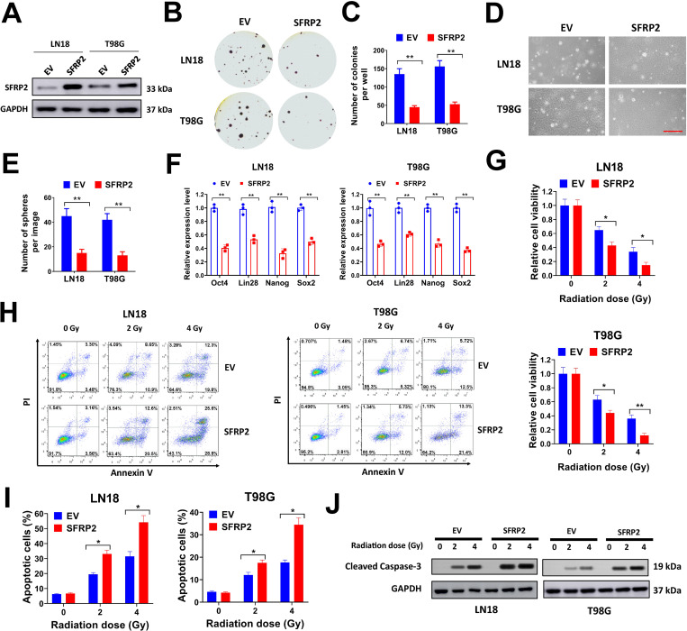
Fig 3. Forced SFRP2 expression suppresses soft agar colony formation, cancer stemness and radioresistance of glioma cells.
A, LN18 and T98G cells were infected with SFRP2 expression lentivirus or empty vector (EV) control, then protein expression of SFRP2 was evaluated by western blot. B-C, LN18 and T98G cells were infected with SFRP2 expression lentivirus or EV control, then seeded in 6-well plates at 8000/well for soft agar assay. Representative plates (B) and average number of colonies per well (C) were shown. D-E, LN18 and T98G cells were infected with SFRP2 expression lentivirus or EV control, then seeded in 6-well plates at 3000/well for sphere formation assay. Representative images (D) and average number of spheres per image (E) were shown. F, expression of Oct4, Lin28, Nanog and Sox2 in LN18 and T98G cells infected with SFRP2 expression lentivirus or EV control was evaluated by qRT-PCR. G, LN18 and T98G cells were infected with SFRP2 expression lentivirus or EV control, then seeded in 96-well plates (3000/well) and treated with a single dose of 2 or 4 Gy X-ray irradiation. Cell viability was tested at 72 h after irradiation. H-J, LN18 and T98G cells infected with SFRP2 expression lentivirus or EV control were seeded in 6-well plates (1 × 10⁶/well) and treated with a single dose of 2 or 4 Gy X-ray irradiation. Then cells were used for flow cytometry (H-I) or western blot (J) at 24 h after the irradiation. Percentages of Annexin-V positive cells (I) were shown. *P< 0.05, **P< 0.001.

