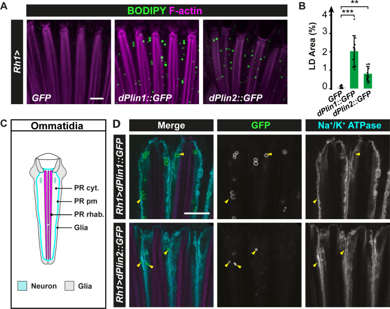
Fig 1. dPlin1 and dPlin2 promote LD accumulation in Drosophila photoreceptor neurons.
(A) LD staining of whole-mount retinas from flies expressing GFP (control), dPlin1::GFP, or dPlin2::GFP in photoreceptor neurons (Rh1-GAL4). LDs are in green (BODIPY) and photoreceptor rhabdomeres are in magenta (phalloidin-rhodamine). Scale bar, 10 μm. (B) Quantification of LD area expressed as % of total retinal area. Data are from the images shown in (A). Mean ± SD. *** p<0.001 by ANOVA with Tukey’s HSD test. (C) Diagram of a longitudinal section of one ommatidium. Each ommatidium is composed of 8 photoreceptor neurons delimitated by a Na+/K+ ATPase positive plasma membrane (cyan), each containing one rhabdomere (magenta), and surrounded by 9 glial cells (also known as retinal pigment cells; light gray). (D) Immunostaining of whole-mount retinas from flies expressing dPlin1::GFP or dPlin2::GFP in photoreceptor neurons (Rh1-GAL4). Photoreceptor plasma membranes are in cyan (anti-Na+/K+ ATPase) and rhabdomeres are in magenta (phalloidin-rhodamine). dPlin1 and dPlin2 are visible as ring shapes in the photoreceptor cytoplasm (yellow arrowheads). Scale bar, 10 μm.

