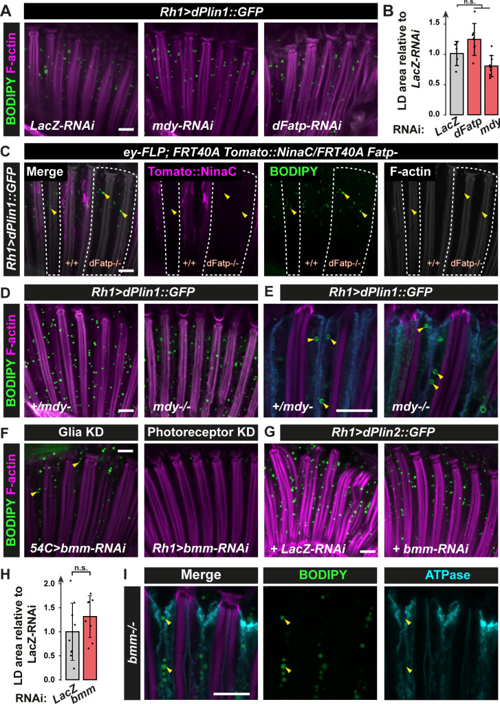
Fig 2. dPlin1-induced LDs in photoreceptor neurons do not require canonical enzymes involved in TG synthesis (dFatp, Mdy) or degradation (Bmm).
(A) LD staining of whole-mount retina from flies with photoreceptor neuron-specific (Rh1-GAL4) expression of dPlin1::GFP and LacZ-RNAi (control), dFatp-RNAi, or mdy-RNAi. LDs are in green (BODIPY) and photoreceptor rhabdomeres are in magenta (phalloidin-rhodamine). Scale bar, 10 μm. (B) Quantification of LD area from the images shown in (A). Mean ± SD. n.s., not significant by ANOVA. (C) LD staining of whole-mount retinas from flipase mediated FRT dFatp[k10307] mutant clone in conjunction with expression of dPlin1::GFP in photoreceptor (Rh1-GAL4). LDs are in green (BODIPY), wild type photoreceptors are in magenta (FRT40A-Tomato[ninaC]) and rhabdomeres are in grey (phalloidin-rhodamine). LDs are visible in photoreceptors lacking dFatp (mutant clone surrounded by dashed line). Scale bar, 10 μm. (D, E) whole-mount retinas from mdy[QX25] heterozygous and homozygous flies expressing dPlin1::GFP in photoreceptor (Rh1-GAL4). (D) LDs are in green (BODIPY), and rhabdomeres are in magenta (phalloidin-rhodamine). (E) Photoreceptor plasma membranes are in cyan (anti-Na+/K+ ATPase) and rhabdomeres are in magenta (phalloidin-rhodamine). (F) LD staining of whole-mount retinas from flies expressing bmm-RNAi in photoreceptor (Rh1-GAL4) or retina glia (54C-GAL4). LDs are in green (BODIPY), and rhabdomeres are in magenta (phalloidin-rhodamine). Scale bar, 10 μm. (G) LD staining of whole-mount retinas from flies expressing dPlin2::GFP in conjunction with RNAi targeting LacZ or bmm in photoreceptor (Rh1-GAL4). LDs are in green (BODIPY), and rhabdomeres are in magenta (phalloidin-rhodamine). Scale bar, 10 μm. (H) Quantification of LD area from the images shown in (G). Mean ± SD. n.s., not significant by t-test. (I) Immunostaining of whole-mount retinas from homozygous bmm mutant flies. Photoreceptor plasma membranes are in cyan (anti-Na+/K+ ATPase) and rhabdomeres are in magenta (phalloidin-rhodamine). Scale bar, 10 μm.

