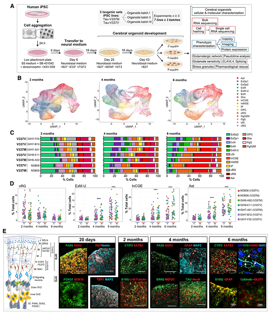
Figure 1. Cerebral organoids exhibit similar differentiation patterns as developing fetal brains.

(A) Experiment summary schematic.
(B) UMAP of scRNA-seq data at 2, 4, and 6 months by cell type. Ast, astrocytes; ExDp1, excitatory deep layer 1; ExDp2, excitatory deep layer 2; ExM, maturing excitatory; ExM-U, maturing excitatory upper enriched; ExN, newborn excitatory; Glia, unspecified glia/non-neuronal cells; InCGE, interneurons caudal ganglionic eminence; InMGE, interneurons medial ganglionic eminence; IP, intermediate progenitors; OPC, oligodendrocyte precursor cells; oRG, outer radial glia; PgG2M, cycling progenitors (G2/M phase); PgS, cycling progenitors (S phase); UN, unspecified neurons; vRG, ventricular radial glia.
(C) Cell type proportions (%) per line at 2, 4, and 6 months.
(D) Cell type proportions (%) for individual organoids over time. Linear model, ***p < 0.001.
(E) Schematic of organoid maturation and neural cell layering (left) and marker visualization (right). Confirmation of dorsal forebrain progenitors at 20 days: PAX6, SOX2, FOXG1, and Nestin; proliferation marker Ki67; absence of SOX10. 2 months: increased MAP2ab, β-III-tubulin neurons; deep-layer glutamatergic neurons TBR1, BCL11B/CTIP2; early glia S100β; few upper layer neurons SATB2+. 4–6 months: few progenitors (SOX2 and PAX6); deep- and upper-layer neurons (BRN2, MEF2C, and SATB2); GFAP+ Ast and Calbindin+ interneurons; robust tau and NeuN; expression of vGLUT1+ and pre- and post-synaptic markers SYN1 and HOMER1, respectively; white arrows indicate adjacent boutons. Scale bars, 100 μm unless otherwise indicated.
See also Figure S1.
