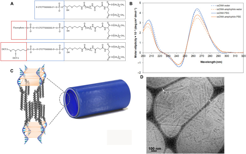
Fig. 1. Ten nucleotide ssDNA-amphiphiles form parallel G-quadruplexes and self-assemble into hollow nanotubes.
(A) Chemical structures of the 10-nt ssDNA-amphiphiles. (B) CD spectra in Milli-Q water and PBS of the free 10-nt ssDNA and the ssDNA-amphiphiles. (C) Schematic representation of the nanotubes and the ssDNA-amphiphiles in the nanotubes, not drawn to scale. ssDNA is shown in blue, and the hydrophobic tails are shown in gray. The light orange represents G-quartet planes. (D) Cryo-TEM image of ssDNA nanotubes. The white arrows point to nanotubes that are viewed end-on, demonstrating their hollow nature.

